Highlight
Sandra Heskamp et al. have published a novel non-invasive imaging technique in Cancer Research.
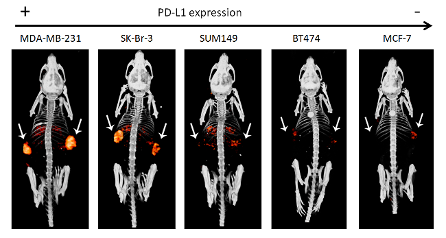
Programmed death ligand 1 (PD-L1) is an import coinhibitory molecule in the anti-cancer immune response. By up-regulating PD-L1, tumor cells are capable of escaping immune recognition and attack. Antibodies that block the interaction between PD-1 and PD-L1 have shown impressive antitumor activity. However, not all patients respond to this type of treatment. Imaging of PD-L1 expression can be used to select patients who are most likely to benefit from anti-PD-1/PD-L1 targeted therapy. In this project, we have developed a novel non-invasive imaging technique to determine tumor PD-L1 expression and accessibility using radiolabeled anti-PD-L1 antibodies and microSPECT/CT imaging. With imaging technique, we can discriminate between tumors with high and low PD-L1 expression levels. The figure shows typical examples of microSPECT/CT images of mice with different types of human breast cancer xenografts. The tumor PD-L1 expression levels increase from left to right To read the full article, please click here.