Research
Intraoperative radiodetection and fluorescence
For many types of cancer, surgical resection is the best or only chance for cure. Incomplete excision of tumor tissue however, negatively affects the prognosis of the patient. In many cases, the presence of residual tumor tissue at the resection margins is a direct predictor of patient survival. To detect tumors or positive resection margins during surgery, visualization techniques like intraoperative molecular imaging can be applied.
To identify tumor tissue specific tumor-targeting radiotracers can be used that can be detected during surgery using a gamma probe. In addition, intraoperative fluorescence imaging allows accurate real-time tumor delineation. These techniques may be used separately depending on the clinical situation, but also a powerful synergy can be achieved by combining radiotracers for the detection of tumor tissue, and fluorescent tracers for subsequent accurate delineation and image-guided resection of tumors.
In my research group, we are working on the development of new tracers to show the potential of these molecular image-guided surgery approaches for the detection of tumor lesions. We have already shown the potential of intraoperative multimodal imaging in renal cell cancer patients (https://pubmed.ncbi.nlm.nih.gov/29721070/) and colorectal cancer patients (https://pubmed.ncbi.nlm.nih.gov/35551444/), and the value of radioguided surgery in prostate cancer patients (https://pubmed.ncbi.nlm.nih.gov/38176721/). In the future, real-time molecular imaging during surgery might become standard of care in many surgical procedures and significantly enhance surgical outcome.
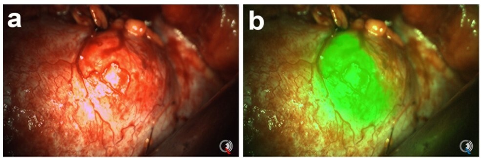
Intraoperative view of a kidney tumor (left) and fluorescence imaging of the tumor tissue in green (right). Adapted from https://pubmed.ncbi.nlm.nih.gov/29721070/.
Researchers involved:
